No products in the cart.

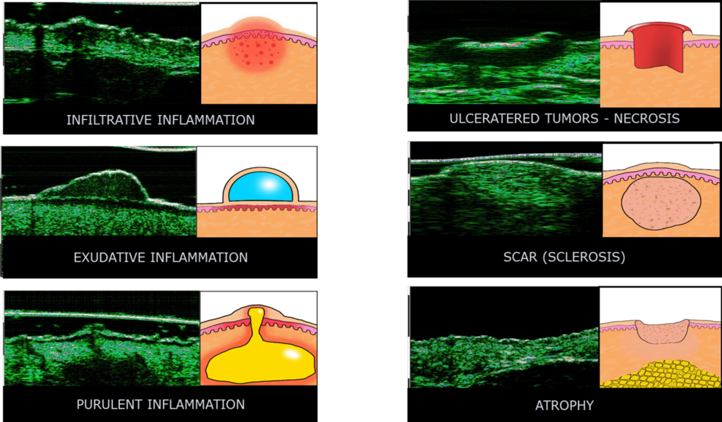
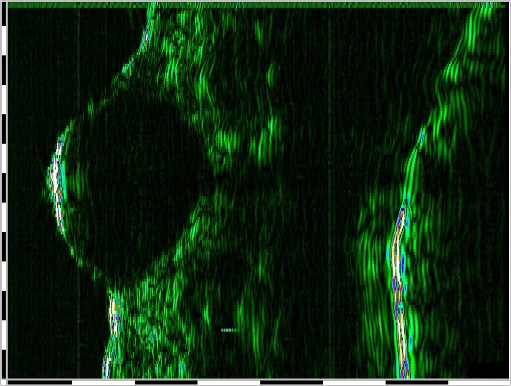
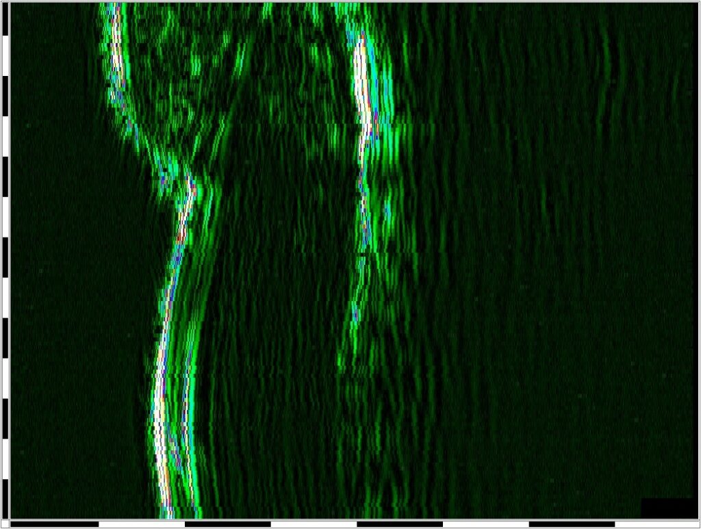
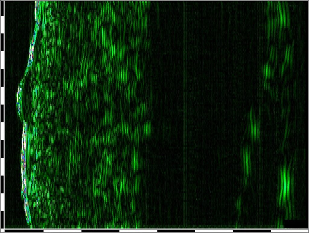
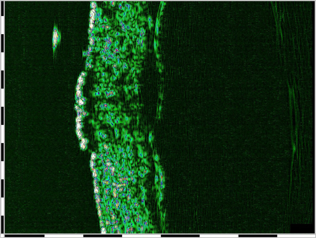
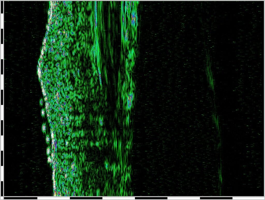
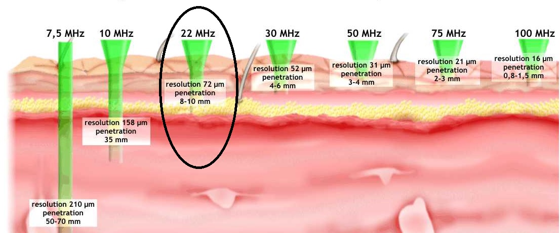
22 MHz high-frequency ultrasound imaging
Allows high-resolution visualization up to approximately 6–8 mm deep into the skin.
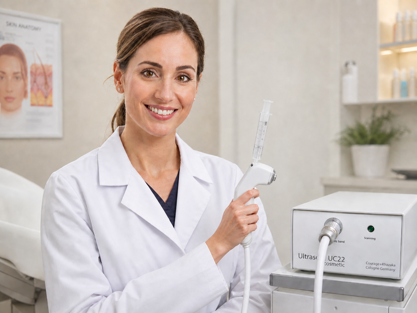
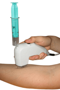
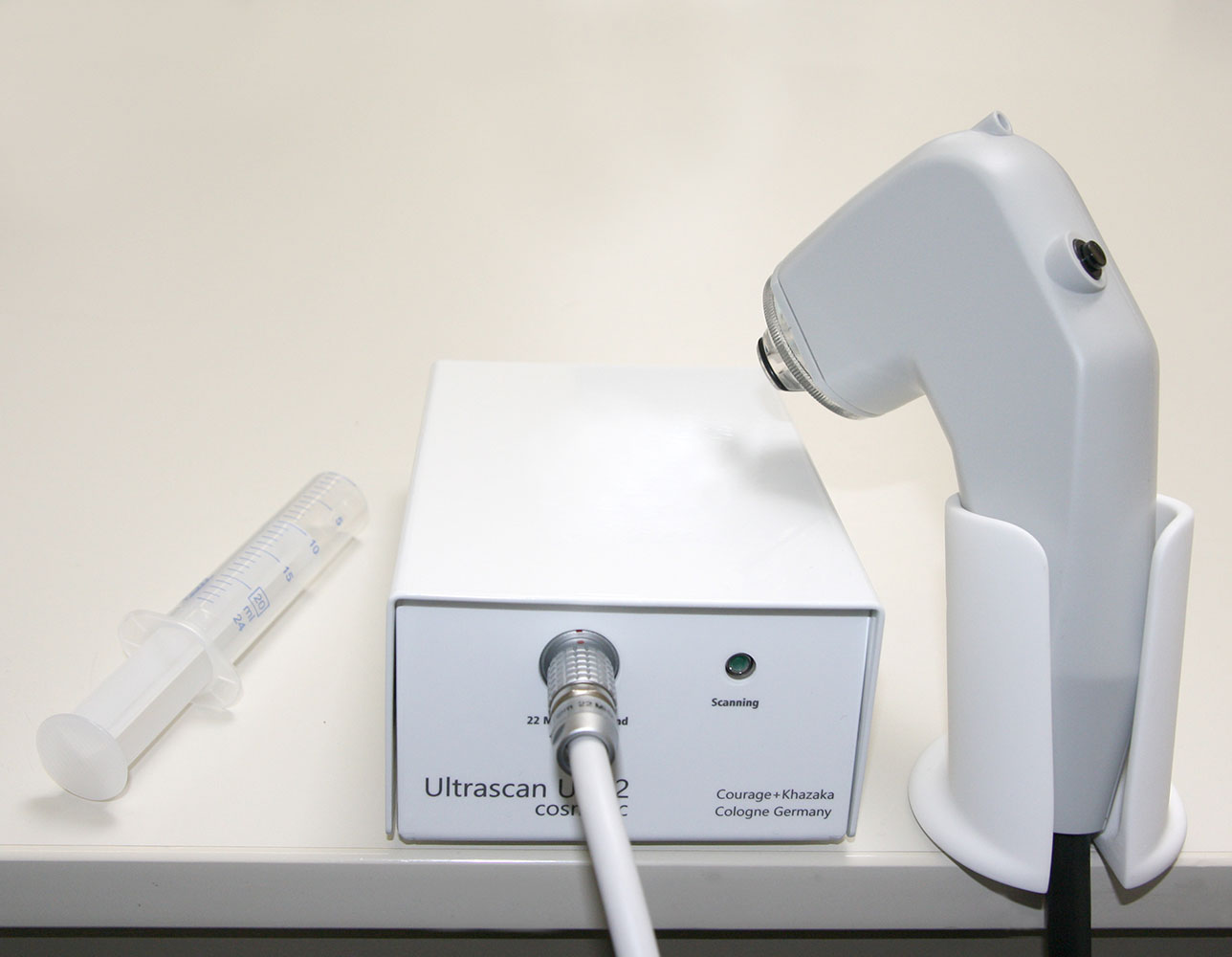
Automatic linear scanning
The probe is easy to handle and performs scans automatically, with the transducer moving linearly over the skin without requiring manual movement by the user.
Water-based scanning
Only water is needed to receive a scan, avoiding messy gel application. Foils are not required but can be used optionally.
Continuous live scan capability
Provides continuous live scans, not just one single image per measurement, with video capture of multiple images per B-scan.
Automatic skin thickness and density measurement
Measures skin thickness in µm and skin density as ultrasound density percentage or colour value, for the total image and separately for the epidermis/entrance echo and dermis.
Advanced measurement tools
Supports measurement of depth, width, line length, and area, including freehand-drawn areas. Measurements of up to 6 areas per image can be saved.
Phase correction function
Flattens the image to help determine skin thickness as accurately as possible.
Adjustable viewing depth
Viewing depth can be changed from 8 mm to 6.4 mm or 4 mm to better visualize details in upper skin layers without changing resolution.
Multiple viewing modes
Supports B-Scan, A-Scan, Sum-A, and ScanLoop viewing modes.
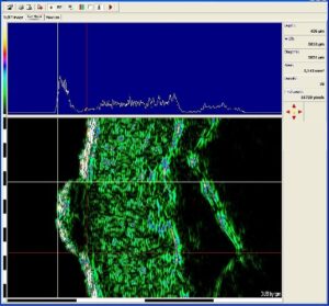
Image export and software compatibility
B-scan images can be exported as image files for use in other applications. The DUB SkinScanner software is compatible with Windows operating systems.
Easy cleaning
The probe head can be cleaned easily after each measurement.
Skin aging and sun damage assessment
Helps evaluate structural changes in the skin related to aging, photoaging, and long-term sun exposure.
Tumour and pre-surgical evaluation
Supports assessment of tumour depth, including applications related to Mohs surgery and pre-surgical planning.
Skin thickness and tissue analysis
Measures skin thickness and helps visualize tissue structure beneath the skin surface, including tissue beneath wounds.
Wound treatment monitoring
Useful for monitoring wound healing, tissue recovery, and changes below the wound surface.
Laser and aesthetic treatment follow-up
Supports before-and-after evaluation of laser treatments, aesthetic skin procedures, and cosmetic interventions.
Cosmetic research and efficacy testing
Provides objective imaging data for product development, claim support, and evaluation of cosmetic treatment effects.
Osteoporosis risk assessment research
Can be used in research applications where skin ultrasound parameters may contribute to osteoporosis risk assessment studies.